About to Mri Front View Of Massive Medial Femoral Condyle Cartilage Defect XxI0 D76noc
Looking for Mri Front View Of Massive Medial Femoral Condyle Cartilage Defect XxI0 D76noc details? We've compiled comprehensive information, latest updates, and exclusive insights for Mri Front View Of Massive Medial Femoral Condyle Cartilage Defect XxI0 D76noc. Discover the complete Details breakdown, history, and detailed profile.
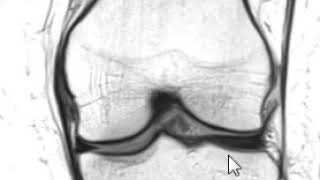
I created the Active Life Orthopedics Guides to help the people I can't see in my practice — practical guidance on recovering from ... Knee pain? A common cause of knee pain is a meniscus tear. Click the link below and request access—I'll approve it for you shortly! In this video Prof. Bellemans explains how to read a knee
Key Details
Explore the primary sources for Mri Front View Of Massive Medial Femoral Condyle Cartilage Defect XxI0 D76noc.
Recent Updates
Stay updated on Mri Front View Of Massive Medial Femoral Condyle Cartilage Defect XxI0 D76noc's newest achievements.
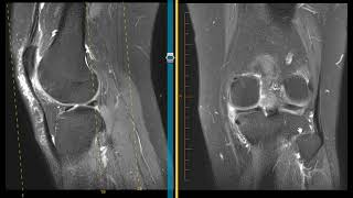
Ultrasonographic examination of grade 4 osteochondral defect in the femoral condyle,
Ultrasound of a High Grade/Full Thickness Medial Sided Quadriceps Tendon Tear in a 30 Year Old Male
How to read a knee MRI?
Detailed Analysis
Data is compiled from public records and verified media reports.
Last Updated: June 18, 2026
Future Outlook
For 2026, Mri Front View Of Massive Medial Femoral Condyle Cartilage Defect XxI0 D76noc remains one of the most talked-about information profiles. Check back for the latest updates.
Disclaimer: Disclaimer: Details details are based on publicly available data, media reports, and general analysis. Actual facts may vary.